The complex auditory disorder known as otosclerosis involves a localized, abnormal process of bone remodeling within the temporal bone, specifically affecting the bony labyrinth that houses the delicate structures of the inner and middle ear. This condition is not merely a form of hardening, as the name suggests, but rather a biphasic process. It begins with an initial, highly vascular, and soft phase called otospongiosis, where bone is actively resorbed by specialized cells. This is then followed by a second phase where the soft, spongy tissue is replaced by dense, disorganized sclerotic bone. The most clinically significant manifestation of this process occurs when this abnormal bone growth targets the stapes, the smallest of the three middle ear ossicles. When the footplate of the stapes, which sits in the oval window leading to the inner ear, becomes stiffened or “fixed” by this sclerotic bone, it can no longer transmit sound vibrations effectively, resulting in progressive hearing loss. This subtle yet relentless progression necessitates a detailed understanding of its signs for effective intervention.
The Most Frequent Chief Complaint of Patients with Clinical Otosclerosis is Hearing Loss
The primary and most frequent symptom that drives patients to seek medical consultation is a progressive hearing loss. This auditory decline typically begins in early adulthood, often presenting between the ages of 15 and 45, and tends to worsen gradually over many years. While it often starts in one ear, it eventually progresses to involve both ears in approximately 70% of individuals, though the degree of loss may not be symmetrical. Patients often initially report a specific difficulty with low-frequency sounds, such as whispers, or struggling to follow conversations in quiet settings. Interestingly, some patients report Paracusis of Willis, a paradoxical phenomenon where they perceive better hearing clarity in noisy environments; this is theorized to occur because the conductive loss filters out some background noise, effectively improving the signal-to-noise ratio of the human voice. This counterintuitive symptom, while not exclusive to otosclerosis, is highly suggestive of a problem in the sound conduction pathway.
Patients often initially report a specific difficulty with low-frequency sounds, such as whispers
Beyond the pure conductive hearing impairment, individuals with otosclerosis frequently experience tinnitus, described as a ringing, roaring, buzzing, or hissing sound in the ears or head. Tinnitus can be a significant source of distress, and its severity may unfortunately increase as the underlying bone disease progresses. Though less common than hearing loss and tinnitus, a number of patients will also complain of dizziness or issues with balance, which can become more pronounced if the otosclerotic focus extends beyond the middle ear to involve the cochlea, leading to a mixed form of hearing loss, incorporating a sensorineural component. These combined symptoms highlight that the disease is not confined to simple mechanics but involves the entire acoustic system.
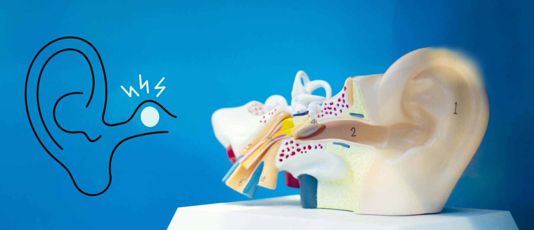
The Classic Radiographic Sign of Otosclerosis is a Radiolucency
The diagnostic process for otosclerosis relies heavily on a combination of patient history, physical examination, and objective audiological and imaging tests. A physical examination using an otoscope is often unremarkable, with the eardrum appearing normal, which helps rule out common middle ear infections or fluid accumulation. However, in about 10% of patients with actively progressing disease, a reddish blush or hue may be visible through the eardrum on the cochlear promontory, known as the Schwartze sign, which is indicative of increased vascularity in the active, spongiotic phase of the bone process. Definitive diagnosis, particularly when surgery is being considered, relies on specialized imaging. High-resolution Computed Tomography (CT) of the temporal bones remains the gold standard for visualizing the bony abnormalities. The classic radiographic sign of otosclerosis is a radiolucency—an area that appears less dense on the scan—at the fissula ante fenestram, the small crevice just anterior to the oval window. This imaging is crucial not only for confirming the presence of the disease but also for assessing the extent of involvement, such as the thickening of the stapes footplate, which is vital for surgical planning.
High-resolution Computed Tomography (CT) of the temporal bones remains the gold standard for visualizing the bony abnormalities
Audiometry, the comprehensive hearing test, provides the necessary functional evidence of the fixation. It typically shows a conductive hearing loss pattern, particularly affecting the low frequencies. A specific finding, often noted in the early stages, is a “Carhart notch”—an artifactual dip in the bone conduction threshold at 2000 Hz. Tympanometry, which measures the mobility of the eardrum, usually yields a Type As curve, indicating a stiffened middle ear system due to the fixed stapes. The combination of these audiological findings with the clinical presentation and, if necessary, the CT results, allows the otolaryngologist to establish a precise diagnosis and prepare a treatment strategy tailored to the individual patient’s needs.
Medical Management with Sodium Fluoride or Bisphosphonates May Help Stabilize Progression
Once a diagnosis of otosclerosis has been established, the treatment path is primarily guided by the severity of the hearing loss, the type of hearing loss, and the patient’s overall health and personal preference. For individuals with mild conductive hearing loss that is not yet significantly impacting daily life, a period of observation or monitoring may be recommended, with periodic audiometric assessments to track the disease progression. Non-surgical treatment options center around two main approaches. The most common non-invasive intervention is the use of hearing aids, which amplify sound to overcome the mechanical barrier created by the fixed stapes. Hearing aids are a safe and effective option, especially for those who either are not candidates for surgery, or prefer to avoid an invasive procedure.
Medical management with sodium fluoride or bisphosphonates may help stabilize progression
In the attempt to slow down the underlying bone remodeling process, medical management has been explored, though its long-term efficacy lacks the robust evidence of large-scale, randomized controlled trials. Some studies have suggested that using sodium fluoride, sometimes combined with calcium and Vitamin D supplements, might help stabilize the progression of the otospongiotic phase and the cochlear component of the disease. The rationale behind this is the potential for fluoride to promote a more stable, less active form of bone. However, this remains a controversial and less frequently utilized primary treatment, usually reserved for patients with active, progressive sensorineural involvement or those for whom surgery is contraindicated.
The Primary Surgical Treatments—Stapedotomy or Stapedectomy—Involve Bypassing or Removing the Immobilized Stapes
For patients whose conductive hearing loss is significant and bothersome, and who have a favorable inner ear reserve—meaning the sensorineural component of their hearing is still relatively good—surgery is typically considered the definitive treatment option. The primary surgical treatments are stapedotomy or stapedectomy, both aimed at restoring the transmission of acoustic energy into the inner ear. The overarching goal is to bypass or remove the immobilized stapes and replace it with a prosthetic device, thereby restoring the continuity of the ossicular chain.
The primary surgical treatments—stapedotomy or stapedectomy—involve bypassing or removing the immobilized stapes
The modern gold standard procedure is the stapedotomy. This technique involves making a small, precise opening (fenestra) in the fixed stapes footplate, often using a laser or a micro-drill, while leaving the majority of the footplate intact. A tiny prosthetic piston is then inserted through this opening, connecting the incus (the middle bone) to the inner ear fluids. This approach is generally preferred over the older stapedectomy, where the entire stapes footplate was removed, because stapedotomy has demonstrated a superior safety profile and is associated with a lower risk of inner ear damage, better preservation of high-frequency hearing, and a reduced incidence of postoperative complications like vertigo. The success rate for significant hearing improvement following stapedotomy, particularly the closure of the air-bone gap, is remarkably high in the hands of experienced surgeons.
Postoperative Vertigo and Taste Disturbance are Relatively Common Immediate Issues
As with any surgical procedure, stapes surgery carries a small but real risk of complications, and patients need to be thoroughly counseled on the potential immediate and long-term sequelae. In the immediate postoperative period, dizziness or vertigo is one of the most common complaints, often lasting for a few hours and sometimes accompanied by nausea or vomiting due to the manipulation of the inner ear. Mild unsteadiness may persist for several days, particularly with quick head movements. Another unique temporary side effect is an altered sense of taste (dysgeusia) on the side of the tongue corresponding to the operated ear. This occurs because the chorda tympani nerve, which carries taste sensation, passes directly through the middle ear and can be stretched or bruised during the surgical approach. In most cases, this taste disturbance resolves spontaneously over several weeks or months.
Postoperative vertigo and taste disturbance are relatively common immediate issues
The most feared, albeit rare, complication is a severe sensorineural hearing loss, or even total deafness, in the operated ear. This can be caused by trauma to the inner ear structures or a perilymphatic fistula—a leak of inner ear fluid. The risk of this significant complication is very low, often cited as less than 1% in high-volume surgical centers. Other potential, though uncommon, complications include facial nerve weakness (usually temporary) and chronic tinnitus that develops or worsens after the procedure. It is imperative that patients adhere strictly to postoperative instructions, which typically involve avoiding activities that increase pressure in the ear, such as flying, heavy lifting, or aggressive nose blowing, for a designated period to ensure optimal healing and reduce the risk of prosthesis displacement or fluid leakage.
The Time Has a Negative Impact on the Results in Form of Air Conduction and Bone Conduction
A critical aspect of understanding otosclerosis and its treatment involves setting realistic expectations regarding the long-term outcomes. While stapedotomy is highly successful in the short term, achieving excellent closure of the air-bone gap, long-term follow-up studies suggest that the auditory gains may not be completely static. Research tracking patients for a decade or more post-surgery has demonstrated a gradual, subtle worsening of both air and bone conduction thresholds over time. This means that while hearing levels typically remain significantly better than they were pre-operatively, there is a slow, progressive decline. This observed worsening is often attributed to the natural aging process of the inner ear (presbycusis), but it can also be a consequence of the underlying cochlear otosclerosis continuing its slow, destructive march.
The time has a negative impact on the results in form of air conduction and bone conduction
Despite this slow, long-term decline, the vast majority of patients maintain a satisfactory level of hearing and continue to benefit significantly from the surgery. However, the data does indicate that some individuals may require a hearing aid as a salvage treatment 10 to 30 years after their initial surgery, suggesting that the operation is not necessarily a permanent cure for the disease’s entirety. This need for long-term monitoring underscores the chronic nature of the otosclerotic process and the necessity for ongoing audiological surveillance.
Tinnitus Was Observed at Presentation in 82 (78.8%) Patients and Spontaneously Resolved or Improved in 51 (62.2%) Patients After Surgery
Tinnitus, as noted, is a pervasive co-symptom of otosclerosis. Its response to surgical intervention is of particular interest to patients. Although the primary goal of stapes surgery is to improve hearing, the secondary benefit of tinnitus reduction is frequently observed. Large case series have indicated that a significant percentage of patients—often over 60%—report that their tinnitus either spontaneously resolved or noticeably improved following the successful restoration of conductive hearing. This outcome is likely due to the normalized input of sound into the inner ear and brain, which diminishes the brain’s compensatory generation of the phantom noise.
Tinnitus was observed at presentation in 82 (78.8%) patients and spontaneously resolved or improved in 51 (62.2%) patients after surgery
However, it is important to emphasize that for a subset of individuals, tinnitus may not change, or it could potentially worsen. Thus, while surgery offers a high probability of alleviating this distressing symptom, it cannot be guaranteed as a tinnitus cure. Patient selection and counseling must therefore be meticulous, ensuring that the primary driver for surgery remains the improvement of the conductive hearing loss, with tinnitus relief considered a positive, though secondary, potential outcome. The intricate relationship between the mechanical action of the middle ear and the resulting neural activity that is perceived as tinnitus highlights the systemic connection between the conductive and sensorineural pathways.